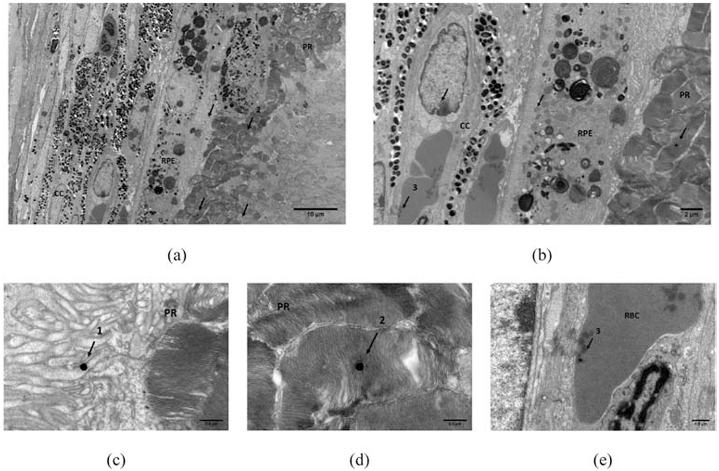
Figure 6:

TEM image of CNV lesion around choroid and RPE-photorecepter showing presence of AuNP (arrows). (a) AuNP present in the PR-RPE and (b) in cc. A high resolution close up of three instances (numbered 1-3 in (a) and(b)) are presented in (c) AuNP’s in RPE, (d) in PR, and (e) in RBC of cc. Scale bars used: (a) = 10 um, (b) = 2 um, and (c)-(e) = 0.5 um. RPE = retinal pigment epithelium, cc = choriocapillaris, PR = photoreceptors, RBC = red blood cell.
