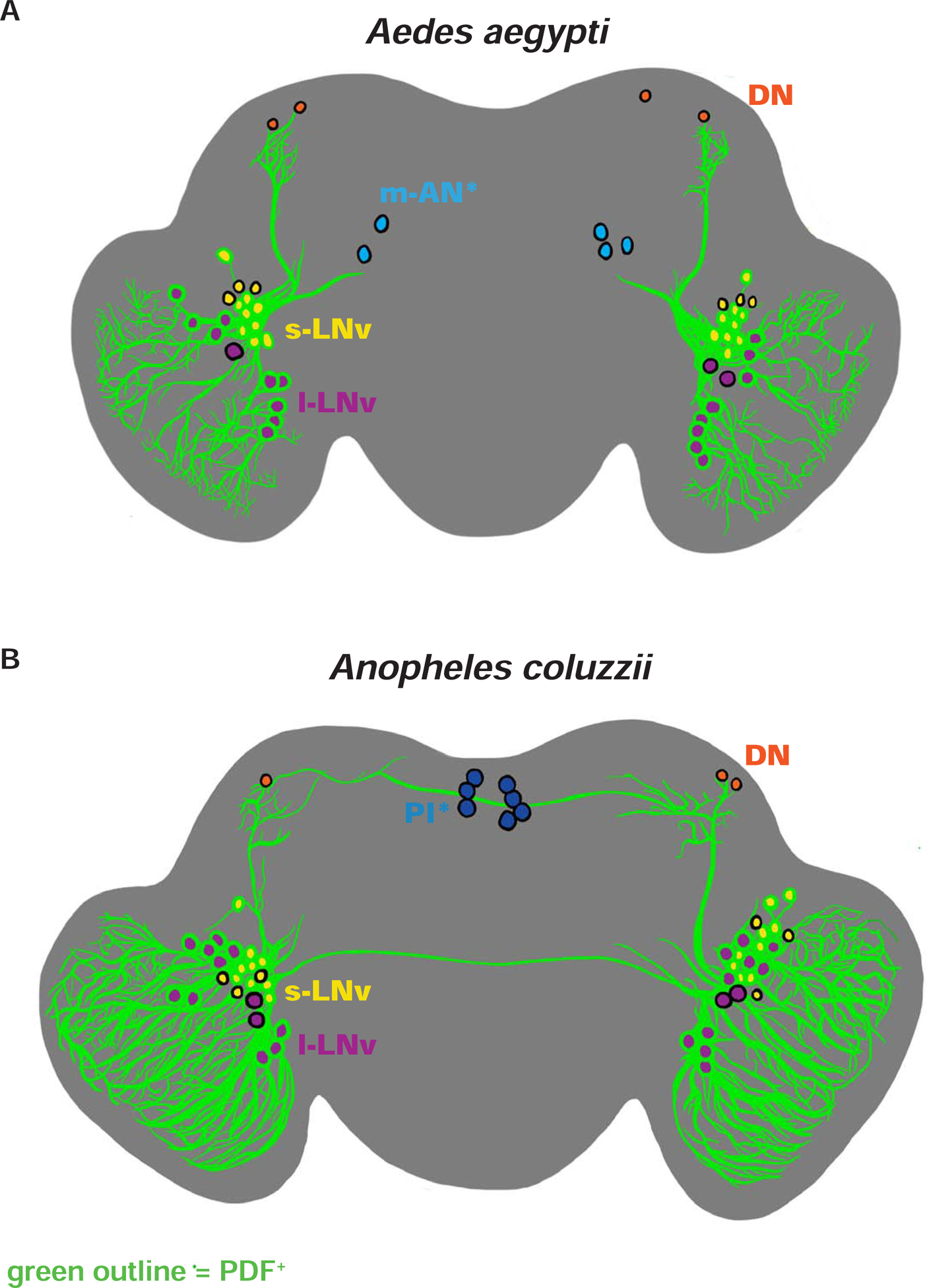
Figure 2. Schematic representation of Aedes aegypti and Anopheles coluzzii circadian neuronal circuits.

(A-B) Illustrations of representative adult female central brains and their neuronal expression of PER and/or PDF with projections depicted in black. Asterisk (*) indicates groups distinct for each species. PDF+ neurons are indicated with green outline. (A) Ae. aegypti s-LNv in yellow, l-LNv in violet, DNs in orange, and m-AN in light blue. (B) An. coluzzii s-LNv in yellow, l-LNv in violet, DNs in orange, and PI neurons in dark blue. See also Figure S2, Figure S3, Figure S4, Table S1, Video S1, and Video S2.
