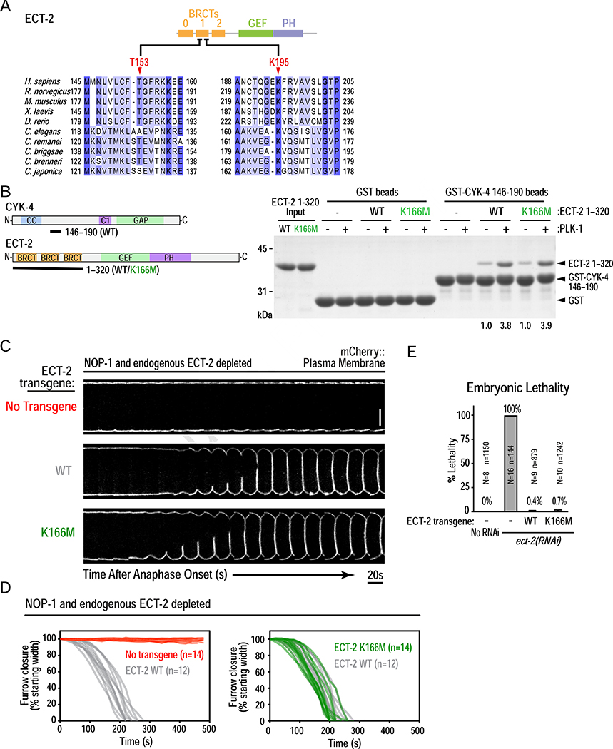
Figure 4. PLK-1 phosphorylated CYK-4 binds to the ECT-2 BRCT repeat domain, but not via its canonical phospho-recognition residues.
(A) Sequence alignment of a section of the human ECT2 BRCT module, with the corresponding region from other vertebrate and nematode sequences. (B) (left) Schematics of proteins used in the pulldown assay, conducted as in Figure 3F. (right) Pulldown results analyzed by SDS-PAGE and Coomassie staining. Numbers below lanes indicate amount of ECT-2 1–320 pulled down, relative to the amount pulled down by unphosphorylated WT CYK-4 fragment. (C) Analysis of furrow ingression for the indicated conditions, done as in Figure 3C. Scale bar, 10 μm. (D) Plots of the kinetics of contractile ring closure in individual embryos for the conditions shown in (C). (E) Plot of embryonic lethality (mean ± SD) for the indicated conditions. N is number of worms and n the number of embryos scored. See also Figure S5.

