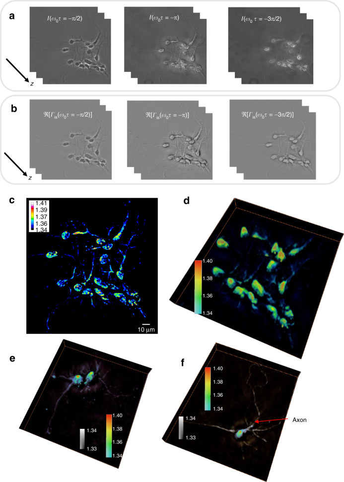
Fig. 4. Wolf phase tomography (WPT) of neurons.
a Three phase-shifted frames of hippocampal neurons (×40/0.75 NA objective). b The real part of the correlation function at three different time lags is solved with Eq. (1). c Refractive index (RI) map of hippocampal neurons. d–f 3D rendering of RI tomograms of hippocampal neurons. e, f Two colormaps are used as indicated to enhance the dendrites and axons. The axon is indicated with a red arrow

