Figures 1–4.
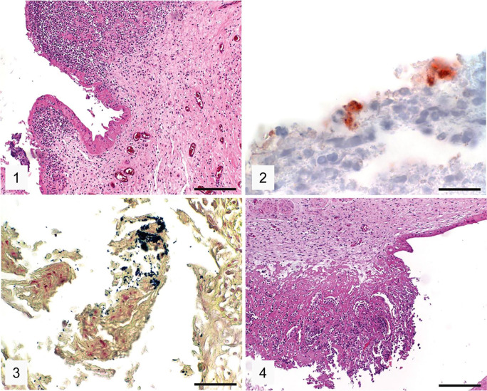
Placenta and umbilicus of the Chlamydia abortus–positive equine abortion case 1. Figure 1. Necrosuppurative placentitis. H&E. Bar = 200 µm. Figure 2. Positive granular intracytoplasmic immunolabeling (red) for Chlamydiaceae in trophoblasts of the placenta. IHC, hematoxylin counterstain. Bar = 20 µm. Figure 3. Presence of gram-positive coccoid bacteria (dark blue) consistent with Rhodococcus equi in the placenta. Gram stain. Bar = 50 µm. Figure 4. Necrosuppurative omphalitis. H&E. Bar = 200 µm.

