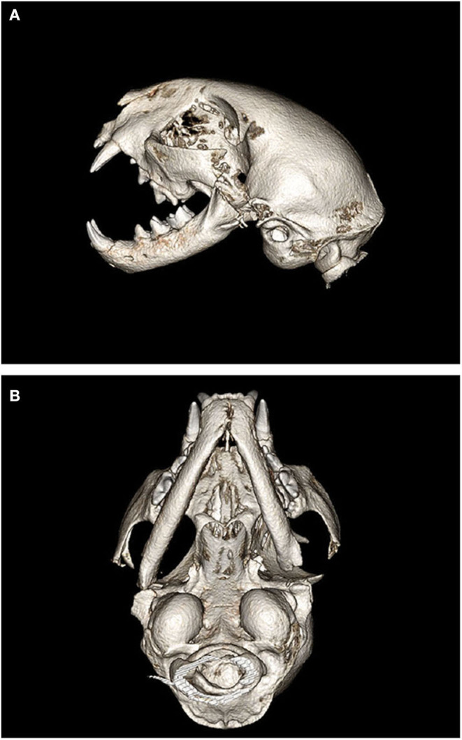
Figure 5.

Postoperative 3D CBCT images of cat #7 after bilateral gap arthroplasty. (A) Left lateral view depicting the ostectomies of the zygoma, coronoid process, condylar process, and mandibular fossa resulting in a “gap.” (B) Ventral view depicting the left sided gap arthroplasty with more conservative ostectomies on the right side.
