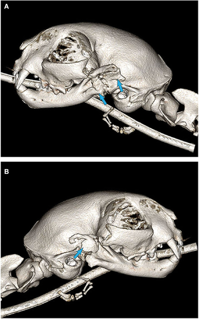
Figure 6.

3D CBCT images of cat #7 6 weeks after initial surgery. (A) Note the substantial new bone formation stemming from the previous ostectomy sites (arrows). (B) New bone formation that is partially organized, at the location of the historically excised coronoid process (arrow).
