FIGURE 3.
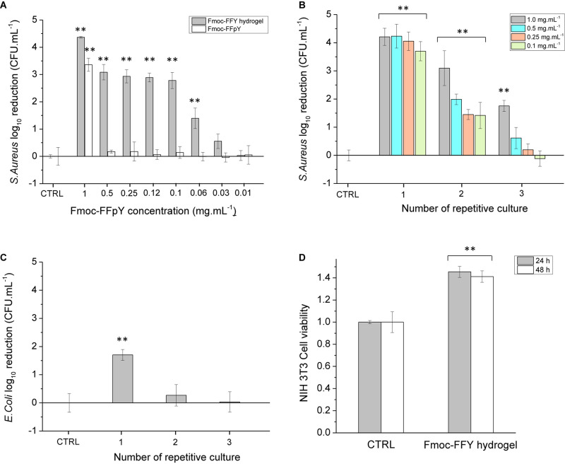
Colony-forming unit (CFU) log10 reduction on planktonic (A) S. aureus cells incubated in the presence of different concentrations of Fmoc-FFpY and of Fmoc-FFY hydrogel, self-assembled with different concentrations of Fmoc-FFpY, (B) S. aureus cells incubated on Fmoc-FFY hydrogel coatings, self-assembled with different concentrations of Fmoc-FFpY on NPs@AP coating and (C) E. coli cells incubated on Fmoc-FFY hydrogel coating, self-assembled with 1 mg.mL– 1 Fmoc-FFpY solution on NPs@AP coating, as a function of the number of repetitive culture. The coating was brought in contact with a fresh pathogen suspension for 24 h. In repetitive culture experiments, the supernatant was removed and replaced every 24 h by a fresh suspension. (D) Cytotoxicity assay of Fmoc-FFY gel through MTT indirect test using NIH 3T3 mouse embryonic fibroblasts cells. Uncoated glass substrates were employed as controls (CTRL). Diagrams include the results from three independent experiments in triplicate and results are shown as mean ± s.e.m. and the ANOVA results at a significance level of **p < 0.01 vs. uncoated glass (negative control, CTRL).

