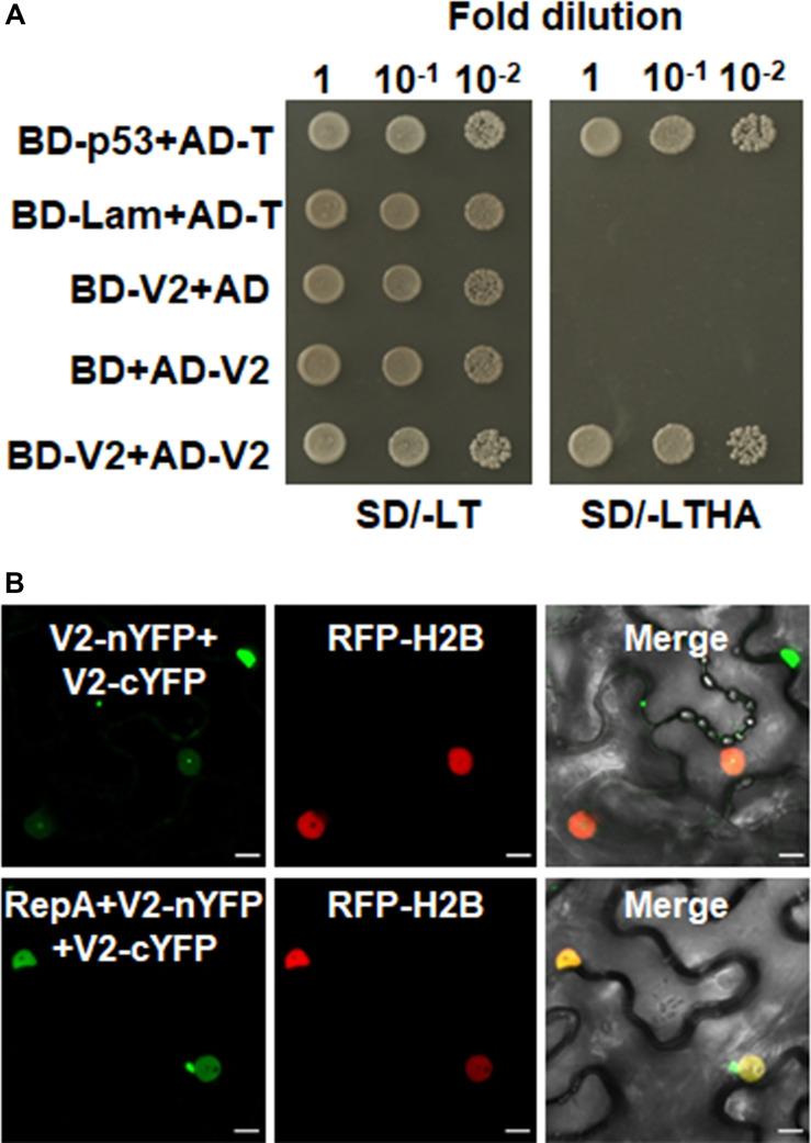
FIGURE 5.
RepA changes the site of V2–V2 self-interaction. (A) Self-interaction of V2 determined by yeast two-hybrid assay. (B) BiFC assays in leave cells of RFP-H2B plants. Self-interaction of V2 in the absence or presence of RepA was examined at 48 hpi using a confocal microscope. YFP signal resulting from interacting protein combinations are indicated as green. RFP-H2B was a marker for the nucleus. Note that the sites of V2–V2 self-interaction was excluded from the nucleolus by RepA. Images are representative of three independent experiments, in each of which at least 20 cells were examined. Scale bars correspond to 10 μm.

