Figure 2.
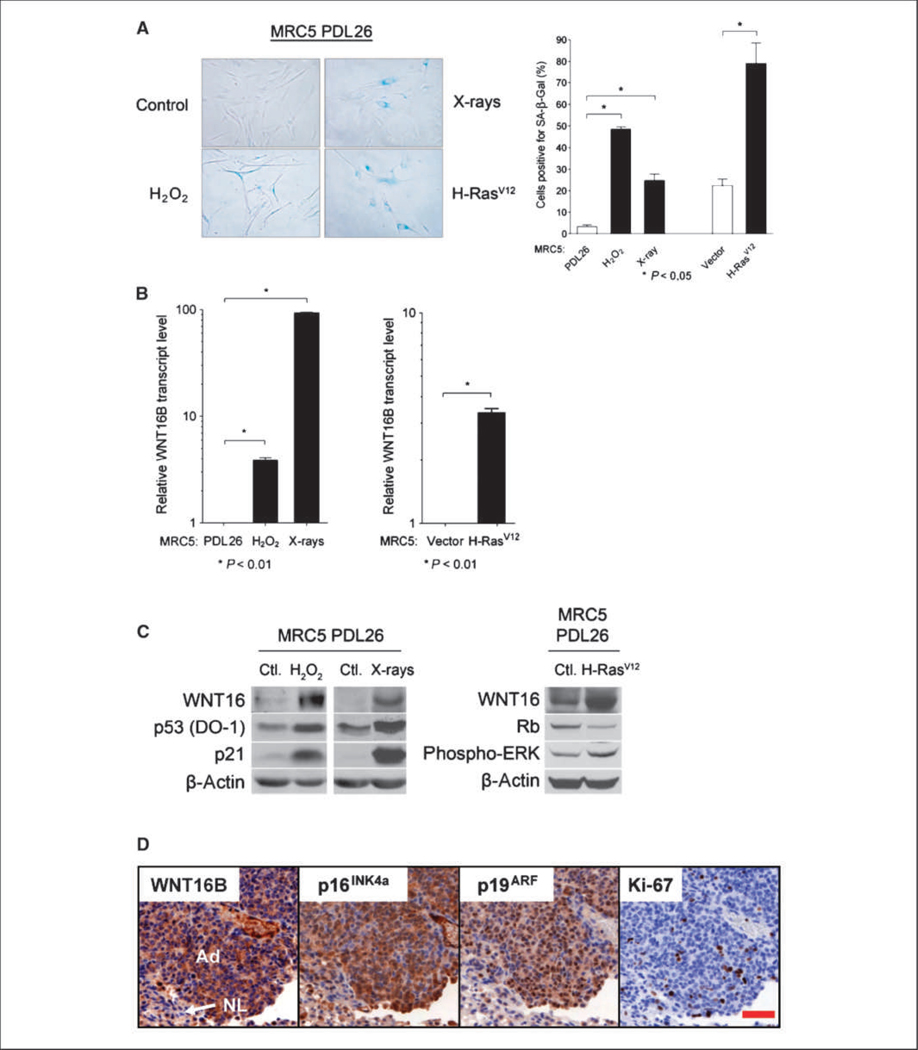
Expression of WNT16B in SIPS and OIS. A, MRC5 fibroblasts were stained for SA-β-Gal expression and quantified. Columns, mean; bars, SD. B, WNT16B mRNA expression was analyzed by qRT-PCR in MRC5 PDL26 fibroblasts exposed to H2O2 or X-rays (left) or infected with a retrovirus coding for H-RasV12 (right). mRNA expression in MRC5 PDL26 fibroblasts was normalized to 1. Columns, mean; bars, SD. C, left and middle, Western blot analysis of WNT16B, p53, and p21 protein expression in MRC5 PDL26 fibroblasts exposed to H2O2 or X-rays; right, WNT16, Rb, and phosphorylated ERK protein expression was analyzed in MRC5 PDL26 fibroblasts infected with a retrovirus coding for H-RasV12. β-Actin was used as a loading control. D, expression of WNT16B during OIS in vivo. Immunohistochemical analysis of WNT16B, p19ARF, p16INK4A, and Ki-67 in serial sections of K-RasV12–induced lung adenomas. Note strong positive staining of WNT16B in the cytoplasm of most tumor cells [adenomas (Ad)] but weak or negative staining in the pocket of nontumoral cells located in the bottom left corner [normal lung (NL)]. Most tumor cells showed nuclear staining of p19ARF often with intense signal in the nucleoli. A fraction of tumor cells gave positive nuclear staining for p16INK4A, whereas the cytoplasmic staining could be nonspecific. Scale bar, 50 μm.

