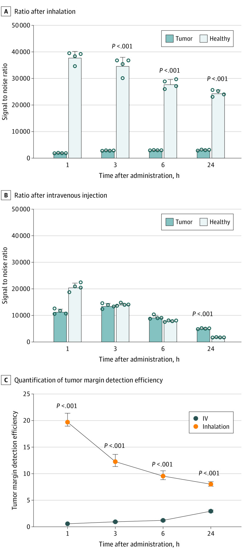
Figure 3. Fluorescent Assessment of Lung Tumor Margin With Inhaled and Intravenously Injected Indocyanine Green (ICG).
Comparison of near-infrared fluorescence signal to noise ratio in tumor and normal tissues at different time points after inhalation (A) or intravenous injection (B) of ICG. Quantification of tumor margin detection efficiency is shown after inhalation and intravenous injection of ICG (C).

