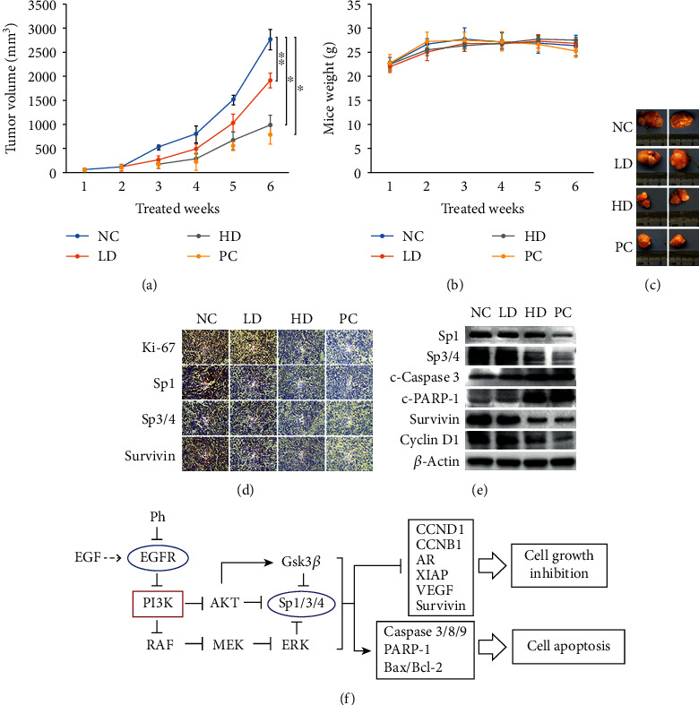
Figure 7.

Phloretin suppressed PC-3 cell xenograft tumor growth and induced PC-3 cell apoptosis in nude mice. The nude mice with PC-3 cell xenograft tumors in the subcutaneous tissue (~30-50 mm3) were treated intraperitoneally with phloretin (low-dose 10 mg/kg and high-dose 50 mg/kg), 1× PBS (as negative control), and 5-FU (as positive control) every two days for 6 weeks. (a) Tumor volumes of the PBS group (negative control), the low-dose phloretin group (low Ph), the high-dose phloretin group (high Ph), and the 5-FU group (positive control) were measured every week, and the average tumor volumes of each group were calculated. ∗P < 0.01, ∗∗P < 0.05. (b) Mouse weights were measured every week and the average mouse weights of four groups were calculated. (c) At the end of the experiment, mice were sacrificed, and subcutaneous tumors were isolated and photographed. (d) The protein levels of Ki-67, Sp1, Sp3/4, and Survivin in tumor tissues were checked by using IHC assay. (e) The protein levels of Sp1, Sp3/4, c-Caspase 3, c-PARP-1, Survivin, and Cyclin D1 in tumor tissues were determined by using western blot assay. (f) Schematic diagram of signal pathways in phloretin-induced cell growth inhibition and apoptosis in human prostate cancer cells. NC: negative control; LD: low dose; HD: high dose; PC: positive control.
