
Figure 3.
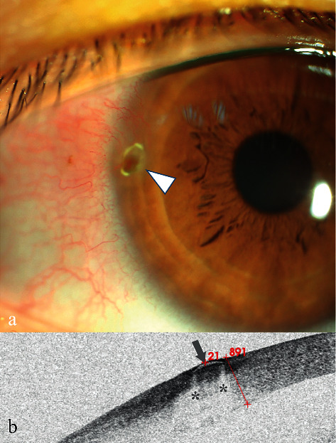
Anterior segment and AS-OCT image for Case 3. (a) Anterior segment photograph: at 9:00 o'clock inside the limbus (arrowhead), a light brown mass was observed without surface fluorescence staining, and fluorescein gathered around the edges. Neovascularization was also seen. (b) AS-OCT image showing that a crescent-shaped low reflective signal (arrowhead) with bilateral marginal zone shadowing (stars) was found 21 μm below the epithelium surface.