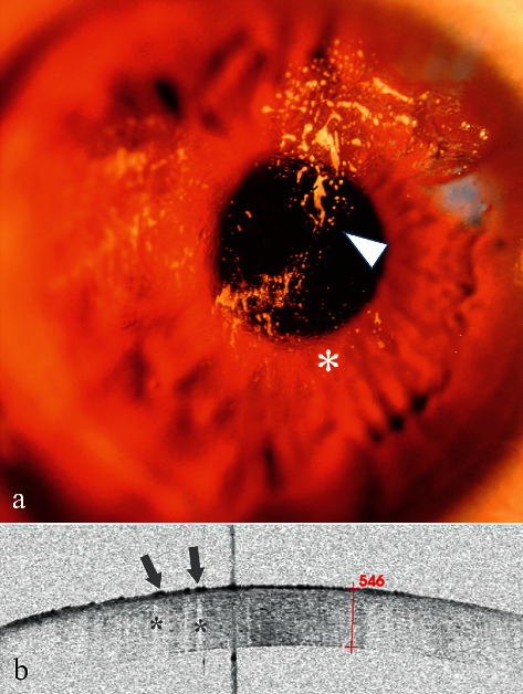
Figure 4.

Anterior segment and AS-OCT image for Case 4. (a) Anterior segment photograph showing multiple cream-like corneal opacities (arrowheads) surrounding a wrinkled transparent cornea. (b) AS-OCT image showed multiple spines (high signals) with blurred boundary (arrows) on the superficial epithelium followed by a central zone shadowing effect (stars). The corneal thickness was 546 μm, within the normal range.
