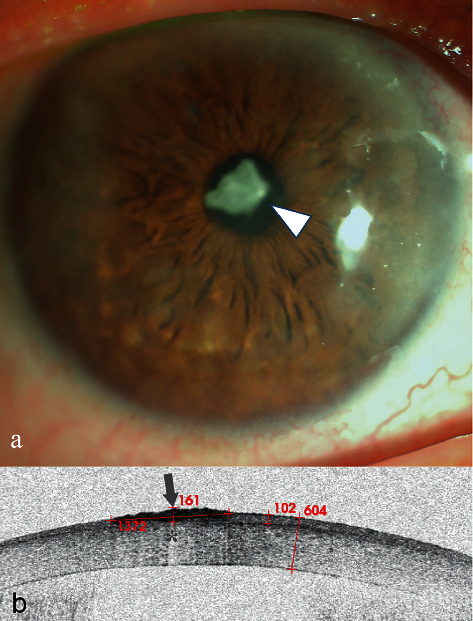
Figure 5.

Anterior segment and AS-OCT image for Case 5. (a) Anterior segment photograph showing a patchy cloud-like haze over the cornea and a cloudy milky lump in front of the pupil (arrowhead). (b) AS-OCT scanning showed there was a lesion of high signal with blurred boundary (arrow) followed by a central zone shadowing effect (star). The lesion had a diameter of 1372 μm. The corneal thickness was 604 μm, and the epithelial thickness was 102 μm. A layer of continuous epithelium tissue was seen on the bottom of the lesion.
