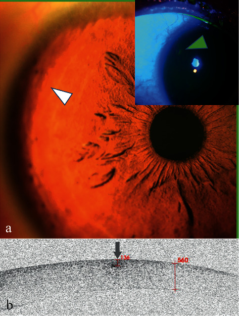
Figure 6.

Anterior segment and AS-OCT image for Case 6. (a) Anterior segment photograph showed a slight bulge at 10:00 o'clock in the peripheral cornea (white arrowhead). Corneal fluorescein staining was negative (arrowhead). (b) AS-OCT scanning showed there was a lesion of high signal with blurred boundary (arrow) without a shadowing effect. The lesion was directly located and was 136 μm beneath the corneal surface.
