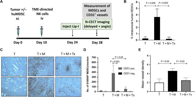
Fig. 2. Intratumoral MDSCs localize to areas of high CD31 vessel density and are eliminated effectively by NKG2D.ζ-modified NK cells.
(A) Experimental schema for evaluating changes in MDSC burden after TME-directed NK cell therapy by flow cytometry, IHC, and nanoparticle contrast–enhanced CT imaging. (B) Intratumoral MDSC burden in tumor-only (T), tumor + MDSC (T + M), and tumor + MDSC + NK cell immunotherapy (T + M + Tx) groups was quantified per group by flow cytometry for CD14+/HLA-DRneg/intracellular S100A9+ cells. (C) Tumors were harvested, sectioned, and analyzed (n = 5 samples per section) for presence of microvasculature by human CD31 immunostaining (brownish red) and of human MDSCs by S100A9 immunostaining (black) on H&E of tissue sections. Shown are two representative sections of tumors inoculated without (T) or with MDSCs (T + M) and tumors with MDSCs after NK cell immunotherapy (T + M + Tx). (D) Number of S100A9+ MDSCs within areas of each tumor section containing CD31+ vessels (CD31 positive) were enumerated and compared to MDSC numbers in areas devoid of CD31+ vessels (CD31 negative). (E) MVD analysis demonstrates a reduction in tumor vascularity after depletion of MDSCs in the NK cell therapy group. Data are presented as means ± SEM (n = 9 to 10 animals per group).

