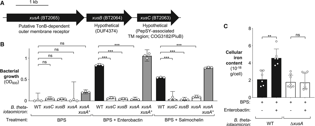
Figure 3: Role of the B. thetaiotaomicron xusABC operon in xenosiderophore uptake in vitro.
(A). Schematic representation of the xusABC operon in B. thetaiotaomicron VPI-5482. (B) The indicated B. thetaiotaomicron strains were cultured in iron-limiting TYG medium supplemented with siderophores for 36 hours. The chelator bathophenanthroline disulfonate (BPS) was added at a concentration of 200 μmol/L. Enterobactin or salmochelin (50% iron saturation) were added at a final concentration of 0.5 μM and 2 μM, respectively. Growth was assessed by measuring optical density (OD600). (C) The B. thetaiotaomicron wild-type strain and an isogenic xusA mutant were cultured in iron-deprived media to exhaust endogenous iron before being subcultured in the presence of iron-laden enterobactin or vehicle. Inductively Coupled Plasma Mass Spectrometry was used to assess cellular iron levels. Bars represent the geometric mean ± 95% confidence interval. **, P < 0.01 ***, P < 0.001; ns, not statistically significant.

