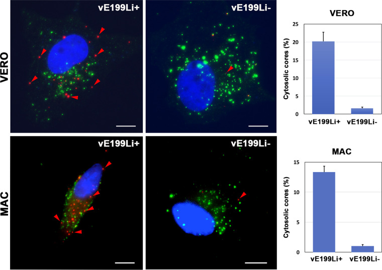
FIG 8.
Protein pE199L is required for core penetration. Vero cells (upper panels) or swine macrophages (bottom panels) were infected with equivalent amounts (25 PFU/cell) of control vE199Li+ and defective vE199Li− virus particles during 3 h at 37°C. After fixation, the cells were permeabilized with saponin and immunostained with antibodies to inner envelope protein p12 (green) and core shell protein p150 (red). Nuclei were stained with Hoechst 33258 (blue). Bars, 5 μm. Note that levels or red punctate p12− p150+ structures, which are interpreted as cytosolic viral cores, are drastically reduced in vE199Li− infections. The proportion of cytosolic cores for each condition and cell type is expressed as a percentage (means ± SD of results from triplicate samples) of the total particles detected per cell (right panels).

