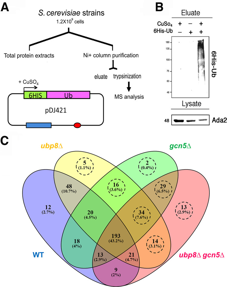
FIG 1.
Expression and purification of His6-Ub proteins in S. cerevisiae. (A) Schematic protocol for the expression of His6Ub proteins in strains containing pDJ421 (41). Red oval, origin of replication; blue rectangle, pCUP promoter (LEU cassette). His6-Ub was expressed in CuSO4, purified through an Ni+ column, and analyzed by MS after trypsinization. (B) Western blot analysis showing the eluate of His6-Ub proteins with respect to the controls hybridized with anti-His6 antibody. Total lysates were probed with anti-Ada2p antibody as an internal standard. (C) Venn diagram of Ub protein distributions found in WT (blue), ubp8Δ (yellow), gcn5Δ (green), and ubp8Δ gcn5Δ (pink) strains. The areas of intersection contain proteins common to different strains. The 113 proteins absent in the WT are highlighted by a dashed circle.

