FIG 3.
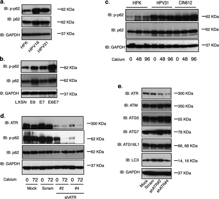
ATR phosphorylates p62 in HPV-positive CIN612 cells. (a) Western blot analysis of p-p62, p62, and GAPDH levels in HFK, HPV16, and HPV31 cells. (b) Western blot analysis of p-p62, p62, and GAPDH levels in HFK cells expressing LXSN vector, HPV31E6, HPV31E7, and HPV31E6/E7. (c) Western blot analysis of p-p62, p62, and GAPDH levels in HFK, HPV31, and HPV31-positive CIN612 cells differentiated in high-calcium media for the indicated times (in hours). (d) Western blot analysis of ATR, p-p62, p62, and GAPDH protein levels in mock, shRNA control, and two different sets of ATR shRNA lentivirus-infected CIN612 cells upon differentiation in high-calcium media for 72 h. (e) Western blot analysis of ATR, ATM, ATG5, ATG7, ATG16L1, LC3, and GAPDH protein levels in mock, shRNA control, and two different sets of ATR shRNA lentivirus-infected CIN612 cells. All results are representative of observations from two or three independent experiments.

