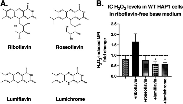
FIG 6.
Mediation of H2O2 entry into HAP1 cells is specific to riboflavin compared to its structural analogs. (A) Chemical structures of riboflavin, roseoflavin, lumiflavin, and lumichrome. (B) HAP1 cells were grown in base DMEMgfp-2 for 24 h, followed by supplementation with 1.063 μM riboflavin (RB), roseoflavin (RF), lumiflavin (LF), or lumichrome (LC) for 16 h, as indicated. Intracellular (IC) levels of H2O2 (determined by Hydrop MFI) in HAP1 cells were measured after exposure to exogenously added 350 μM H2O2 or vehicle. MFI fold change data shown represent MFI of H2O2-treated cells relative to MFI of untreated cells. A fold change value of 1 (dotted line) represents no change. Error bars represent SEM (n = 6 independent experiments). *, P < 0.05 (compared to cells in riboflavin-containing medium [+RB]).

