
FIG 7.
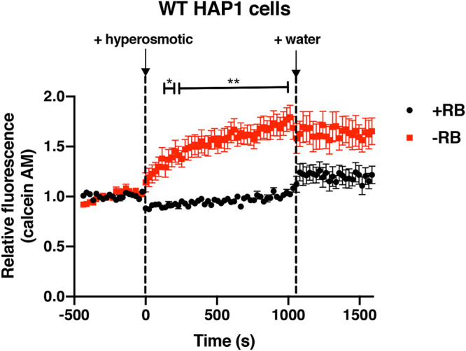
Effects of riboflavin on osmotic regulation in HAP1 cells. WT HAP1 cells were grown in base DMEMgfp-2 supplemented with 1.063 μM riboflavin (+RB) or in base DMEMgfp-2 without riboflavin (−RB) for 48 h. Relative fluorescence levels of calcein AM (a self-quenching dye) were used to determine volume changes upon addition of a hyperosmotic solution (2.5 M NaCl) to cells and then upon addition of water to cells. Error bars represent SEM (n = 12 independent experiments). Two-way ANOVAs were used to determine statistical differences between relative fluorescence values in samples (+RB or −RB) at different time points. *, P < 0.05; **, P < 0.01 (compared to +RB samples).