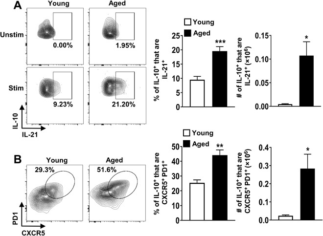
Fig. 2. IL-10–producing FoxP3−CD4+ T cells in aged mice are predominantly Tfh cells.

(A) Splenocytes from young (2 months, n = 6) and aged (18 months, n = 6) C57BL/6 mice were stimulated with P + I, stained with antibodies against TCRβ, CD8, FoxP3, IL-10, and IL-21, and analyzed by flow cytometry. The representative plots and graphs show the frequencies and total numbers of IL-21+ cells originating from FoxP3−IL10+ cells (means ± SEM). Data are representative of at least two independent experiments. (B) Splenocytes from young (2 months, n = 4) and aged (18 months, n = 4) C57BL/6 mice were stimulated as above and stained with antibodies against TCRβ, CD8, FoxP3, CXCR5, PD1, and IL-10 and analyzed by flow cytometry. The representative plots and graphs show the frequencies and total numbers of CXCR5+PD1+ cells originating from FoxP3−IL10+ cells (means ± SEM). *P ≤ 0.05, **P ≤ 0.01, and ***P ≤ 0.001, Student’s t test.
