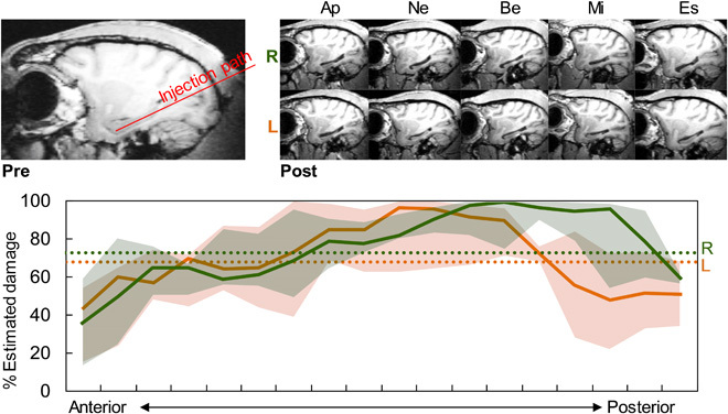
Fig. 1. Excitotoxic injections of N-methyl-d-aspartate produced selective hippocampal lesions.

(Top) Left: Presurgery sagittal MRI showing intact hippocampus and injection path. Right: Postsurgery sagittal MRIs of both right (R) and left (L) hemispheres from five monkeys showing shrunken hippocampi. (Bottom) Percent estimated cell loss for each hemisphere along the anterior-posterior axis. Solid lines are group medians, shaded areas are first and third quartiles, and dotted lines are overall means for each hemisphere.
