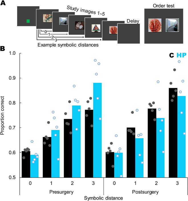
Fig. 3. Hippocampal damage did not reliably impair memory for temporal order.

(A) Trial sequence showing a green start square, five study images, a retention delay, and then a test in which the monkey had to touch the image that had appeared first during study. Example symbolic distances are indicated by arrows. The depicted test shows images 5 and 2, for a symbolic distance of 2. (B) Proportion correct as a function of symbolic distance, surgical time point, and group (control = black bars and filled gray dots, hippocampal = blue bars and open blue dots). Postoperatively, monkeys showed a strong SDE (F3,24 = 84.30, P < 0.001) but there was no difference between the groups in overall accuracy (t8 = 0.67, P = 0.520), no interaction of symbolic distance and group (F3,24 = 0.50, P = 0.689), and no group difference at any individual symbolic distance (all t8 < 1.15, all P > 0.284). Bars represent group means, each dot represents one monkey, and dots are jittered to allow visualization of individual performance. Chance is 0.5. Compare figure 2 of (7). Stimuli images from Flickr under a Creative Commons CC BY 2.0 Generic License.
