Fig. 1.
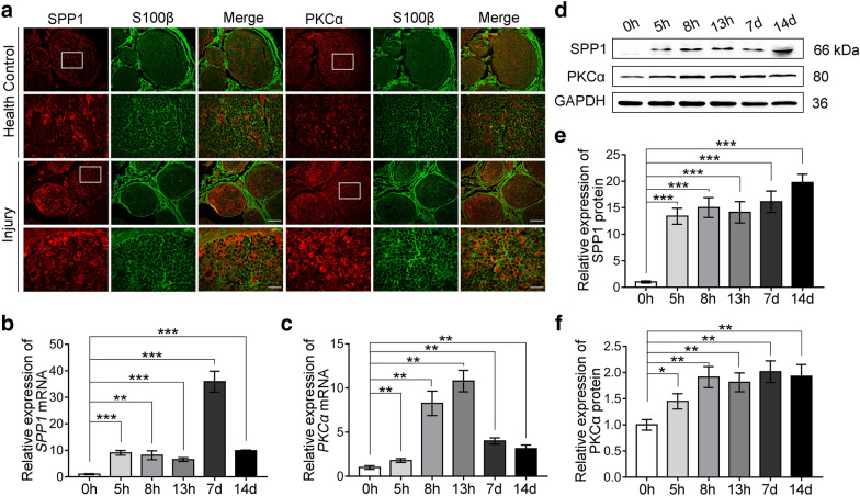
Expression of SPP1 and PKCα was increased in SCs after median nerve injury in the human specimens. a Immunofluorescence staining of SPP1 (red), PKCα (red) and S100β (green) in health median nerve and distal median nerve stumps at 5 days after injury. Scale bar, 200 and 50 μm. b, c Relative mRNA expression of SPP1 (b) and PKCα (c) in injured median nerve at 0, 5, 8, 13 h, 7, and 14 days of six independent samples by real-time qPCR. d–f Western blot analysis of SPP1 and PKCα in injured median nerve at 0, 5, 8, 13 h, 7, and 14 days after injury. Repeated for three-time. d Representative blot results. e, f Quantification of the relative expression of proteins in (d). Data are presented as mean ± SEM. *P < 0.05, **P < 0.01, ***P < 0.001

