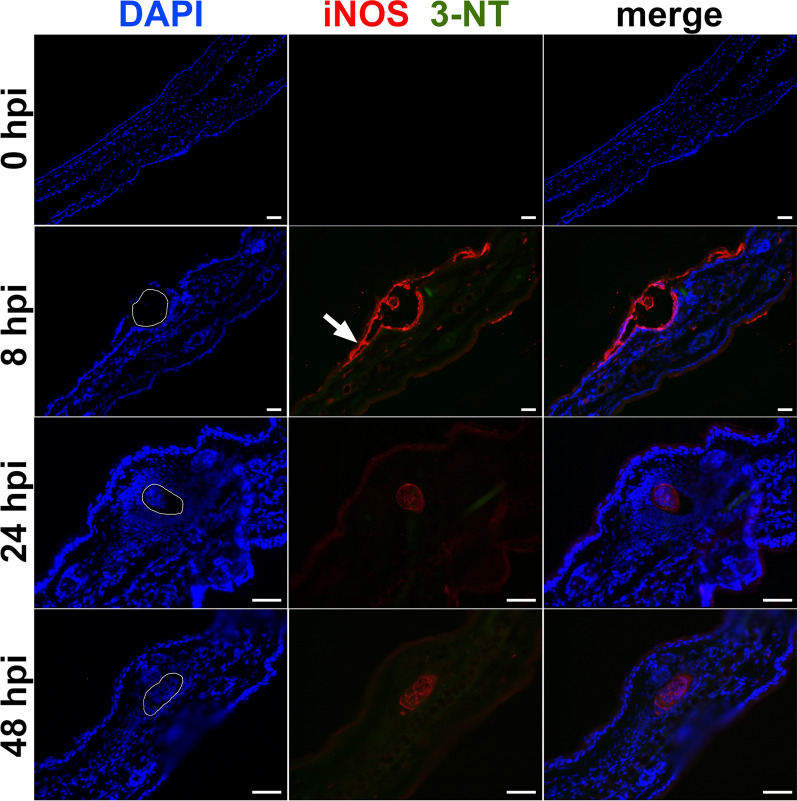
Fig. 1.
Immunolocalization of inducible nitric oxide synthase (iNOS) a 3-nitrotyrosine (3-NT) in pinnae of mice infected with Trichobilharzia regenti. The iNOS signal (red) surrounded the penetrating schistosomula 8 h post-infection (hpi) and spread also in the adjacent epidermal area (white arrow). Leukocytes infiltrated the lesion 24 hpi, but neither iNOS nor 3-NT signals were detected at this or later (48 hpi) time points. The space occupied by schistosomula is marked with white line in DAPI images. Three mice per time point and at least two schistosomula-positive slides per mouse were analyzed. The faint red signal observed 24 and 48 hpi represents a non-specific binding of the rabbit polyclonal antibody to the schistosomula and was observed also in control samples incubated with negative rabbit serum (not shown). Scale-bars: 50 μm

