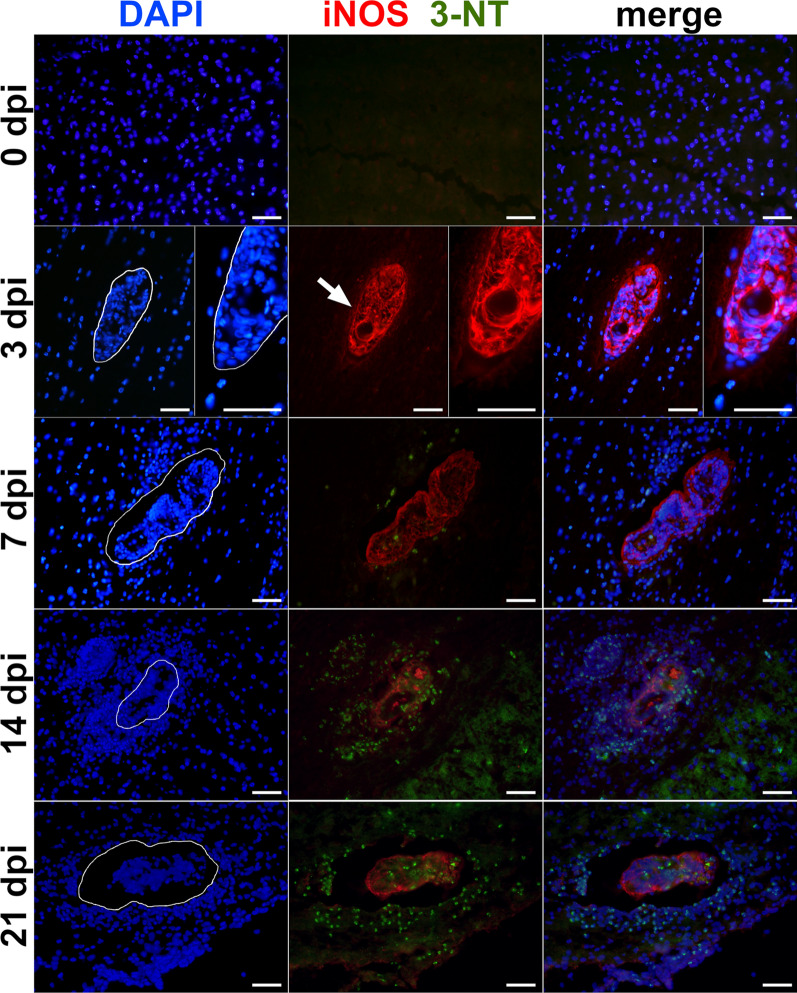
Fig. 2.
Immunolocalization of inducible nitric oxide synthase (iNOS) a 3-nitrotyrosine (3-NT) in the spinal cord of mice infected with Trichobilharzia regenti. The iNOS signal (red, pointed by white arrow) surrounded schistosomula 3 days post-infection (dpi). The signal of 3-NT (green) started to appear 7 dpi and was more frequent 14 and 21 dpi. It was localized within the cells surrounding the schistosomula as well within the schistosomula internal tissues. The space occupied by schistosomula is marked with white line in DAPI images. Three mice per time point and at least two schistosomula-positive slides per mouse were analyzed. Similarly to Fig. 1, the non-specific binding of the rabbit polyclonal anti-iNOS antibody to the schistosomula was observed. Scale-bars: 50 μm

