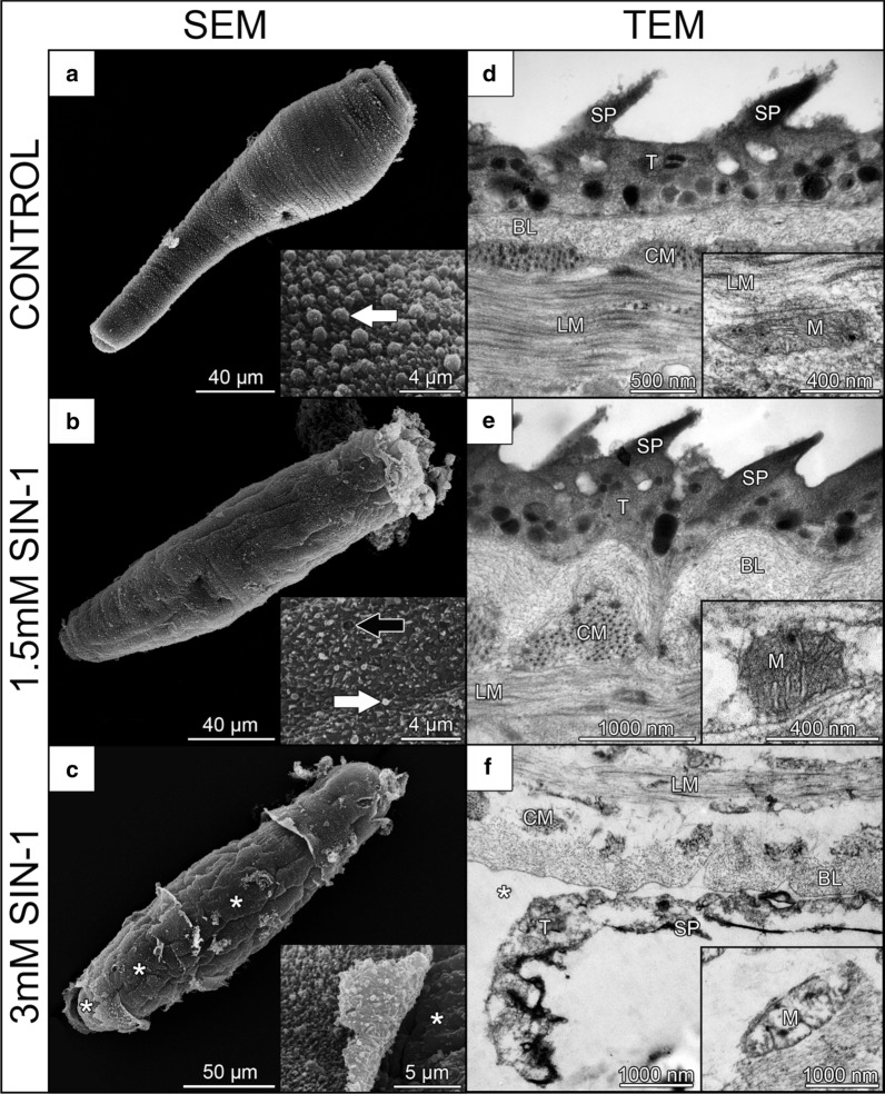
Fig. 6.
Ultrastructural changes of Trichobilharzia regenti schistosomula treated with SIN-1, the donor of peroxynitrite. The schistosomula were treated for 48 h and then examined under scanning (SEM; a-c) and transmission (TEM; d–f) electron microscopy. As revealed by SEM, the tegument of 1.5 mM SIN-1-treated schistosomula had fewer blebs (white arrows) covering the layer of spines than the tegument of the control group. Also, the topography of tegumental spines was changed after 1.5 mM SIN-1 treatment and small holes (black arrow) appeared in the surface (b). No differences were observed between these groups in TEM (d, e) including ultrastructure of mitochondria (insets in d, e). Schistosomula treated with 3 mM SIN-1 had severely destructed tegument partially revealing the basal lamina (white asterisks) (c, f). Subtegumental mitochondria had disrupted inner membranes and cristae (inset in f). Abbreviations: T, tegument; SP, spines; BL, basal lamina; CM, circular muscles; LM, longitudinal muscles; M, mitochondria

