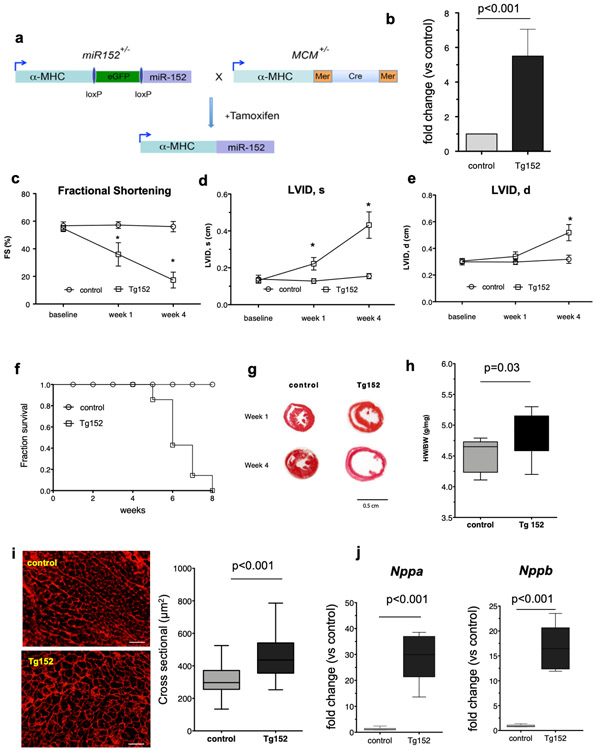
Figure 2. Forced miR-152 expression in cadiomyocytes induces cardiac dilatation and overt cardiac dysfunction in mice.
a) Schematic diagram of the Cre-LoxP–mediated gene-switch strategy to establish transgenic animals with temporally regulated gene overexpression of miR-152 in the myocardium. Tamoxifen-induced gene switch in the hearts of double transgenic animals carrying both miR-152 and MCM alleles. MCM: MerCreMer; a-MHC: α-myosin heavy chain. b) Real-time PCR expression analysis of miR-152 gene expression in the myocardium of bi-trangenic mice (Tg152) and littermate MCM control mice (n=4 per group). Comparison by paired Student’s t-test. c-e) Echocardiography analysis of cardiac function and dimensions of Tg152 mice and MCM control littermates prior to and at 1- and 4-weeks post tamoxifen treatment (n=6 per group at each time point). Data represent mean *P < 0.001 by 2-way ANOVA repeated measures test. f) Survival curve of Tg152 and control animals following tamoxifen administration. g) Representative Hematoxylin-Eosin staining of heart sections at 1 week and 4 weeks post tamoxifen treatment. h) Heart-weight-to-body weight ratios (n=9 animals per group). i) Assessment of cardiomyocyte diameters by wheat germ agglutinin staining (n=250 cardiomyocytes per group). Scale bar = 100 μm. j) Real-time PCR expression analysis of Nppb and Nppa genes in the myocardium of bi-trangenic mice (Tg152) and littermate MCM control mice (n=6 per group). All Box-and-whisker plots show the minimum, the 25th percentile, the median, the 75th percentile, and the maximum. FS=Fractional Shortening; LVID,s=Left ventricular internal diameter at end systole; LVID,d = Left ventricular internal diameter at end diastole.

