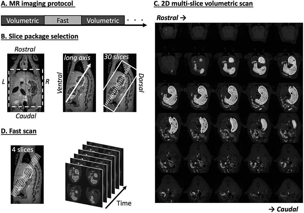
Fig. 1.

Imaging sequence and slice package selection. A. 2D multi-slice volumetric scan (denoted as “Volumetric”) and fast antral motility scan (denoted as “Fast”) were performed interleaved throughout the experiment. B. Two example T2-weighted images (left: coronal view, right: sagittal view) acquired from a multi-slice localizer sequence. An oblique 30-slice package was placed along the long axis of the stomach from the sagittal image. C. Example semi-coronal view images from the 2D multi-slice volumetric scan. D. Fast scan of the antrum with four slices. The position of the slice package was determined from images acquired from the volumetric scan.
