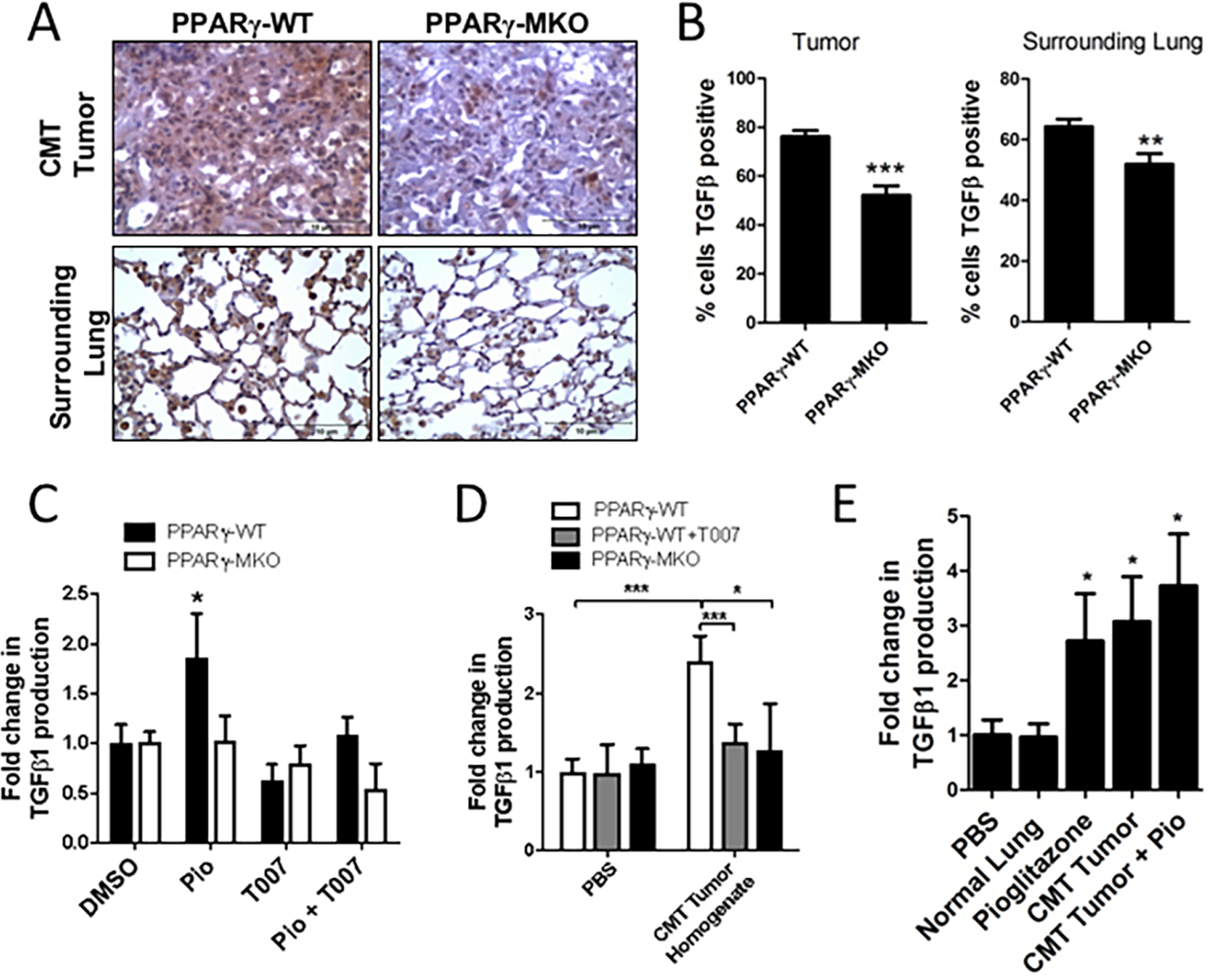
Figure 1: TGF-β1 is produced by myeloid cells in response to PPARγ activation.

(A) PPARγ-WT or PPARγ-MKO mice were orthotopically injected with CMT-luc cells. Tumor bearing lungs were collected 4 weeks later. IHC for TGF-β1 was performed on tissue sections and representative images at 40X magnification are shown. (B) Quantification of TGF-β1 positive cells within tumor and surrounding lung tissue in IHC sections, reported as a percent of the total cells. (C) TGF-β1 released from bone marrow derived macrophages (BMDM) from PPARγ-WT or PPARγ-MKO mice treated with pioglitazone (Pio), PPARγ inhibitor (T007), a combination of both, or vehicle control (DMSO) and normalized to WT DMSO control. (D) TGF-β1 released from PPARγ-WT or PPARγ-MKO BMDM treated with media supplemented with CMT-luc tumor homogenate and normalized to PPARγ-WT PBS control. (E) TGF-β1 released from PPARγ-WT BMDM treated with media supplemented with homogenate from normal lung or lung with CMT-luc tumor (CMT), pioglitazone (Pio) or a combination of tumor homogenate and pioglitazone and normalized to WT vehicle control (*p<0.05, **p<0.01, ***p<0.001).
