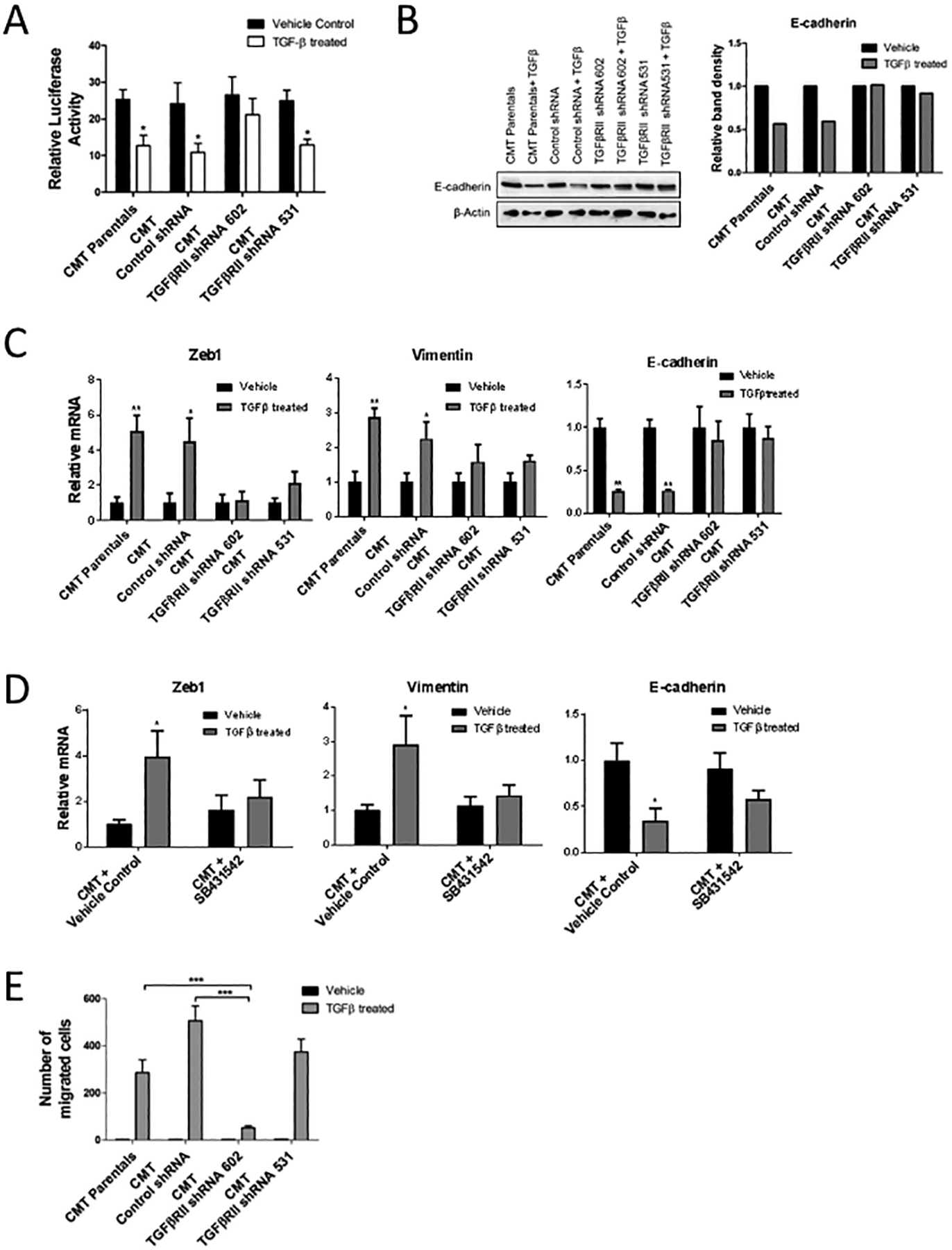
Figure 3: Response of CMT-luc cells treated with TGF-β1 in vitro.

(A) Proliferation of cells treated with TGF-β1 or vehicle control in vitro, measured as change in total luminescence (*p<0.05 for TGF-β1 treated compared to respective vehicle treated controls). (B) Representative western blot for E-cadherin in CMT-luc cells treated with TGF-β1 compared to control with corresponding densitometry analysis reported as relative band intensity. (C) qRT-PCR for Zeb1, Vimentin and E-cadherin in CMT-luc cells post TGF-β1 treatment compared to control. (D) qRT-PCR for E-cadherin, Zeb1, and Vimentin in CMT-luc cells post TGF-β1 treatment combined with the TGFβRI inhibitor SB431542 or vehicle control (DMSO). (E) Number of migrating cells in transwell migration assays using CMT-luc cells treated with TGF-β1 or vehicle control (**p<0.01, ***p<0.001).
