
Figure 4: LLC-luc induce PPARγ dependent myeloid cell TGF-β1 production but do not require TGF-β1 signaling for progression.
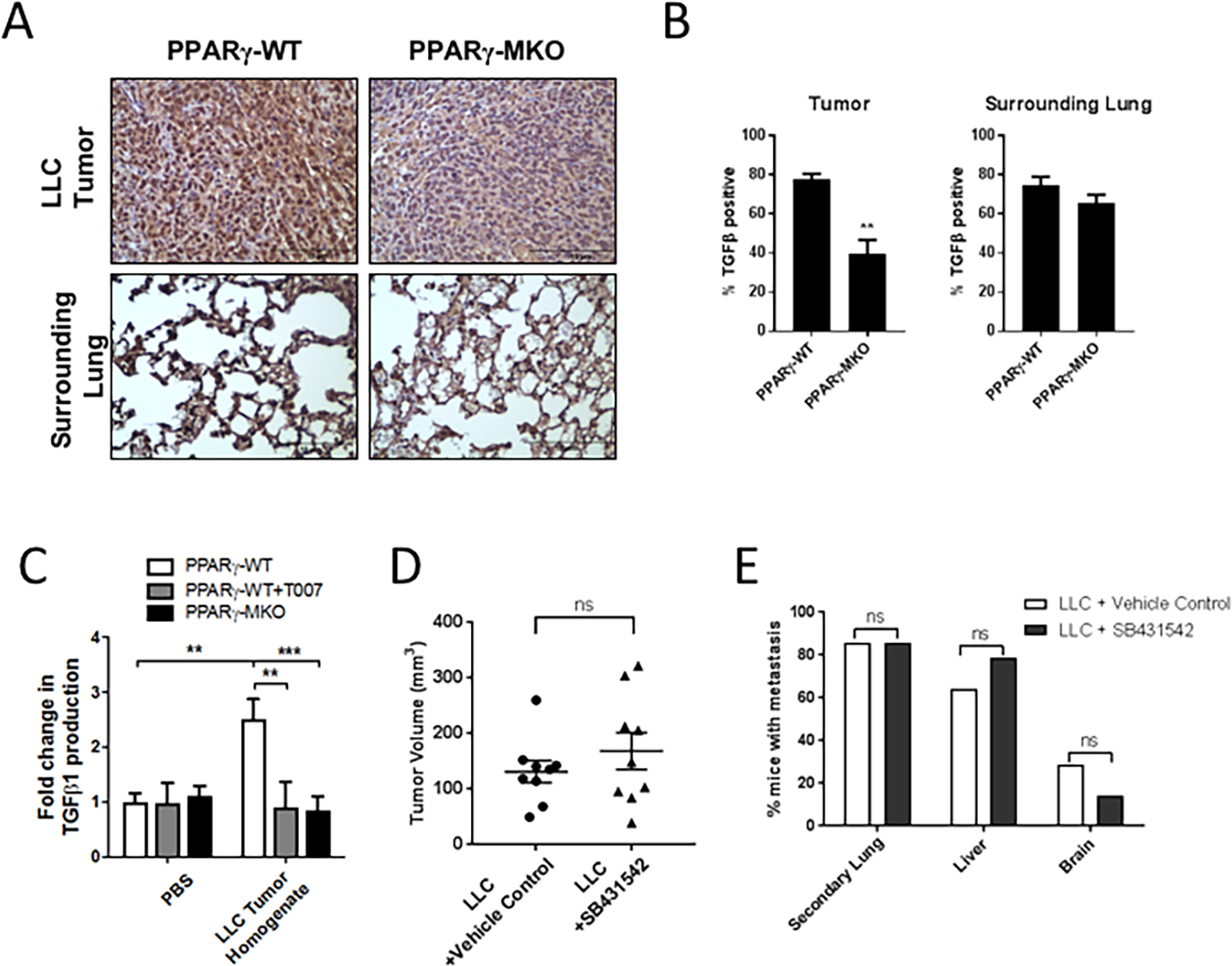
(A) PPARγ-WT or PPARγ-MKO mice were orthotopically injected with LLC-luc cells. Tumor bearing lungs were collected 2.5 weeks later. IHC for TGF-β1 was performed on tissue sections and representative images at 40X magnification are shown. (B) Quantification of TGF-β1 positive cells within tumor and surrounding lung tissue in IHC sections, reported as a percent of the total. (C) TGF-β1 released from PPARγ-WT or PPARγ-MKO BMDM treated with media supplemented with LLC-luc tumor homogenate. (D) WT mice were orthotopically injected with LLC-luc cells and treated with the TGFβRI inhibitor SB431542 or vehicle control (1:1 DMSO:PBS) starting at the time of injection and continuing 5 days a week for 2.5 weeks. There were 8 mice per group injected with LLC-luc cells. Primary tumor size was measured by caliper and reported as the tumor volume in mm3. (E) Incidence of metastasis reported as the percent of mice with metastasis to the other lobes of the lung (secondary lung), liver, or brain as measured by bioluminescence (**p<0.01, ***p<0.001).