
Fig. 2. arid1aa and arid1ab haploinsufficiency enhances MYCN-induced sympathoadrenal lineage hyperplasia by increasing cell proliferation in the IRG.
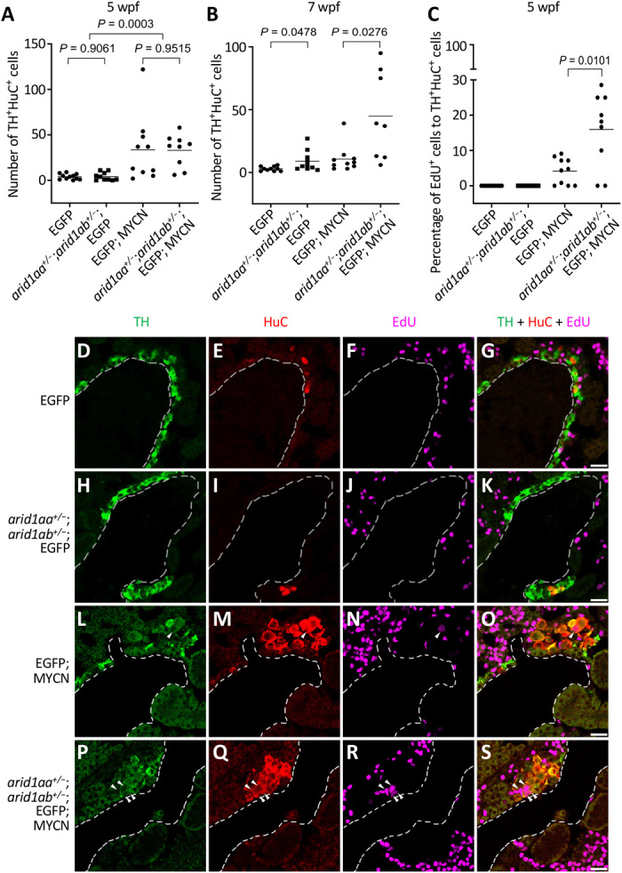
(A and B) Numbers of TH+HuC+ sympathoadrenal cells in the IRG of EGFP, arid1aa+/−;arid1ab+/−;EGFP, EGFP;MYCN, and arid1aa+/−;arid1ab+/−;EGFP;MYCN lines at both 5 wpf (A) and 7 wpf (B). (C) Percentage of EdU-positive TH+HuC+ sympathoadrenal cells in the IRG of the indicated fish lines at 5 wpf. In all plots, each symbol represents the value for an individual fish. Horizontal bars indicate mean values that were compared with the two-tailed unpaired Welch t test. (D to S) Representative coimmunostaining images of TH (green), HuC (red), and EdU (magenta) of the sagittal sections through IRG of EdU-labeled EGFP (D to G), arid1aa+/−;arid1ab+/−;EGFP (H to K), EGFP;MYCN (L to O), and arid1aa+/−;arid1ab+/−;EGFP;MYCN (P to S) lines at 5 wpf. Arrowheads indicate the EdU-labeled proliferating sympathoadrenal cells. Dotted lines indicate the head kidney boundary. Scale bars, 20 μm.