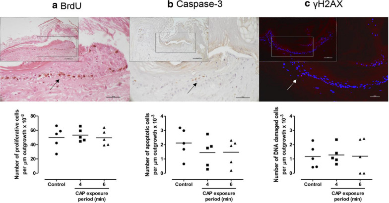
Fig. 6.
Effect of repeated exposure to CAP on ex vivo wound healing. During 2 weeks culture, BWMs were exposed four times (twice weekly) to CAP for 4 or 6 min or not exposed (negative control). Subsequently, the number of proliferative (a), apoptotic (b) and DNA damaged (c) cells per µm of newly formed epidermis (outgrowth) was determined using immunohistochemistry. The arrows indicate the positively-stained cells in the outgrowth of the unexposed BWMs (scale bars: 50 µm). This is also shown at a smaller magnification in the inset (scale bars: 100 µm). Data represent the means of five independent experiments performed in duplicate. No statistically significant differences were measured (Wilcoxon S-R; p > 0.05)

