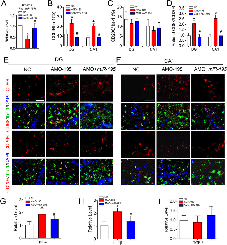
Fig. 4.
Knockdown of miR-195 prime microglial/macrophage polarization to M1 phenotype in rats. a MiR-195 expression was detected by qRT-PCR in the hippocampus of rats following the stereotaxic injection of lenti-pre-AMO-miR-195 and/or lenti-pre-miR-195 into CA1 region. Bars represent the mean ± SD. n = 6. *P < 0.05 vs NC group, #P < 0.05 vs AMO-195. b Changes of miR-195 affect the percentage of CD68 in the Iba-1+cells in hippocampal DG, CA1 region of rats. c Changes of miR-195 has no effects on the percentage of CD206 in the Iba-1+cells in hippocampal DG, CA1 region of rats. d Changes of miR-195 regulate the ratio of CD68/CD206 in the hippocampal DG and CA1 regions. Bars represent the mean ± SD. n = 9 slices from 3 animals per group, *P < 0.05 vs NC group, #P < 0.05 vs AMO-195. e, f Representative images of CD68 and CD206 expression in the Iba-1+ cells of rat hippocampal DG (e) and CA1 (f) region following the stereotaxic injection of lenti-pre-AMO-miR-195 and/or lenti-pre-miR-195 into CA1 region by immunofluorescence staining. The scale bar was 40 μm. g The mRNA expression of TNF-α in the hippocampus of rats following the stereotaxic injection of lenti-pre-AMO-miR-195 and/or lenti-pre-miR-195 into CA1 region. h The mRNA expression of IL-1β in the hippocampus of rats following the stereotaxic injection of lenti-pre-AMO-miR-195 and/or lenti-pre-miR-195 into CA1 region. i The mRNA expression of TGF-β in the hippocampus of rats following the stereotaxic injection of lenti-pre-AMO-miR-195 and/or lenti-pre-miR-195 into CA1 region. Bars represent the mean ± SD. n = 6. *P < 0.05 vs NC group, #P < 0.05 vs AMO-195. All data were analyzed using one-way ANOVA followed by Tukey test

