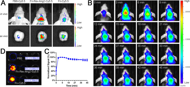
Figure 2.
Fn-Rev-Ang-Cy5.5 crossed the BBB in vivo. (A) Fluorescence imaging of Cy5.5-PBS (left), Fn-Rev-Ang-Cy5.5 (middle), and Fn-Cy5.5 (right) in vivo and ex vivo following the injection of each solution into the tail vein. (B,C) Fluorescence imaging in vivo. Mice were injected i.v. with Fn-Rev-Ang-Cy5.5 and were imaged every 3 min for 45 min (D) Fluorescence imaging showing that the number of Fn and the number of Fn-Rev-Ang-labeled fluorescence were the same.

