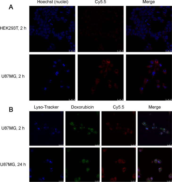
Figure 3.
(A) Confocal imaging of the intracellular uptake of Fn-Rev-Ang-Dox-Cy5.5 in HEK293T cells at 2 h of incubation (top row) and in U87MG cells at 2 h. Scale bar, 25 μm; Hoechst, blue; Cy5.5, red. (B) Confocal imaging of the intracellular uptake of Fn-Rev-Ang-Dox-Cy5.5 in U87MG cells at 2 and 24 h. Scale bar, 25 μm; LysoTracker, blue; Dox, green; Cy5.5, red.

