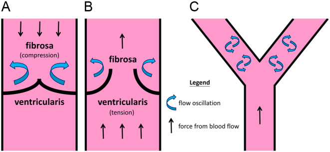
Figure 5.

Regions of oscillatory blood flow in valve vs in blood vessel bifurcations. (A and B) Illustrate oscillatory flow on the leaflet’s fibrosa side and the distribution of flexural compression (fibrosa) and tensile stresses (ventricularis) during valve open and closed states, respectively. (C) Depicts a straight segment of vasculature that experiences laminar flow. Oscillatory flow is formed at bifurcation sites downstream of the straight segment. It is observed that regions of flow oscillations are most vulnerable to plaque formation or calcification (76, 77).

 This work is licensed under a