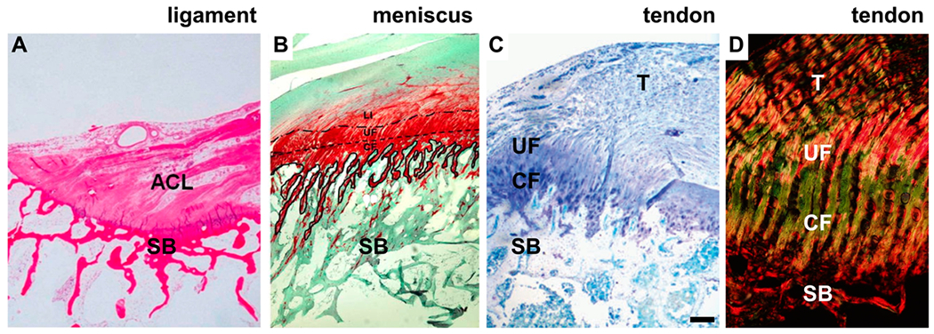
Figure 1.

Enthesis of the anterior cruciate ligament (ACL), meniscus, and rotator cuff tendon, shown histologically. SB = subchondral bone, CF = calcified fibrocartilage, UF = unmineralized fibrocartilage, and T = tendon. (A) Histology of the ACL enthesis at the tibial attachment, stained using hematoxylin and eosin.43 (B) Histology of the meniscal enthesis, stained using Safranin O,44 highlights the fibrocartilage region rich in proteoglycan in red. The dashed line in B indicates the tidemark between SB and CF. (C and D) Histology of the supraspinatus enthesis of the rotator cuff tendon, stained using (C) toluidine blue45 for enhanced metachromatic stain of the fibrocartilage (purple color) and (D) picrosirius red for enhanced birefringence under polarized light. Scale bar in C = 200 μm. The image in A was adapted with permission from ref 43. Copyright 2014 Sage. The image in B was adapted with permission from ref 44. Copyright 2008 Springer. The image in C was modified with permission from 45. Copyright 2011 International Bone and Mineral Society.
