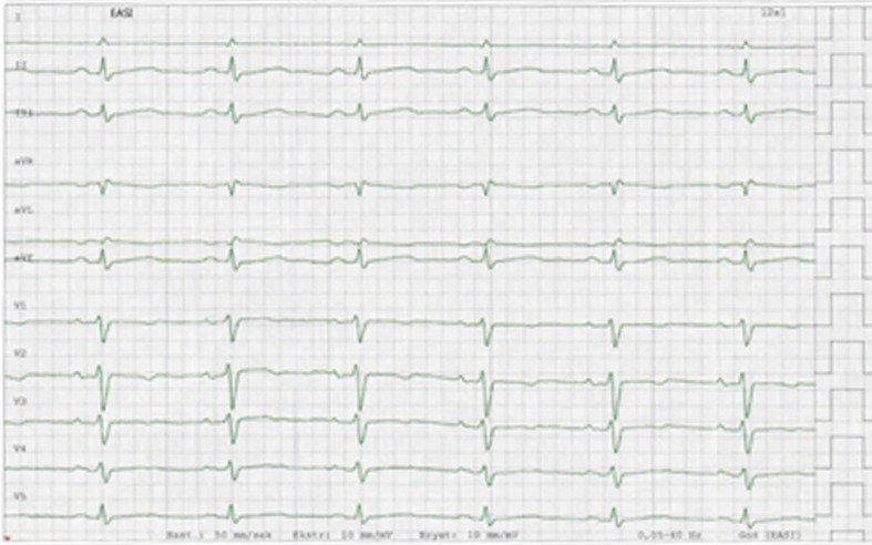
Figure 1.
EASI ECG representative for the course of disease. The ECG was recorded on day 14. Lead V6 is lacking in the stored version. There is insignificant ST-elevation in inferior leads and T-wave inversion in precordial leads. Furthermore, low voltage findings are present with peak-to-peak QRS amplitude less than 5 mm in the standard leads and 10 mm in the precordial leads (V5 and V6). Similar findings were seen in the printed 12-lead ECGs recorded at admittance to hospital and during the hospital stay.

