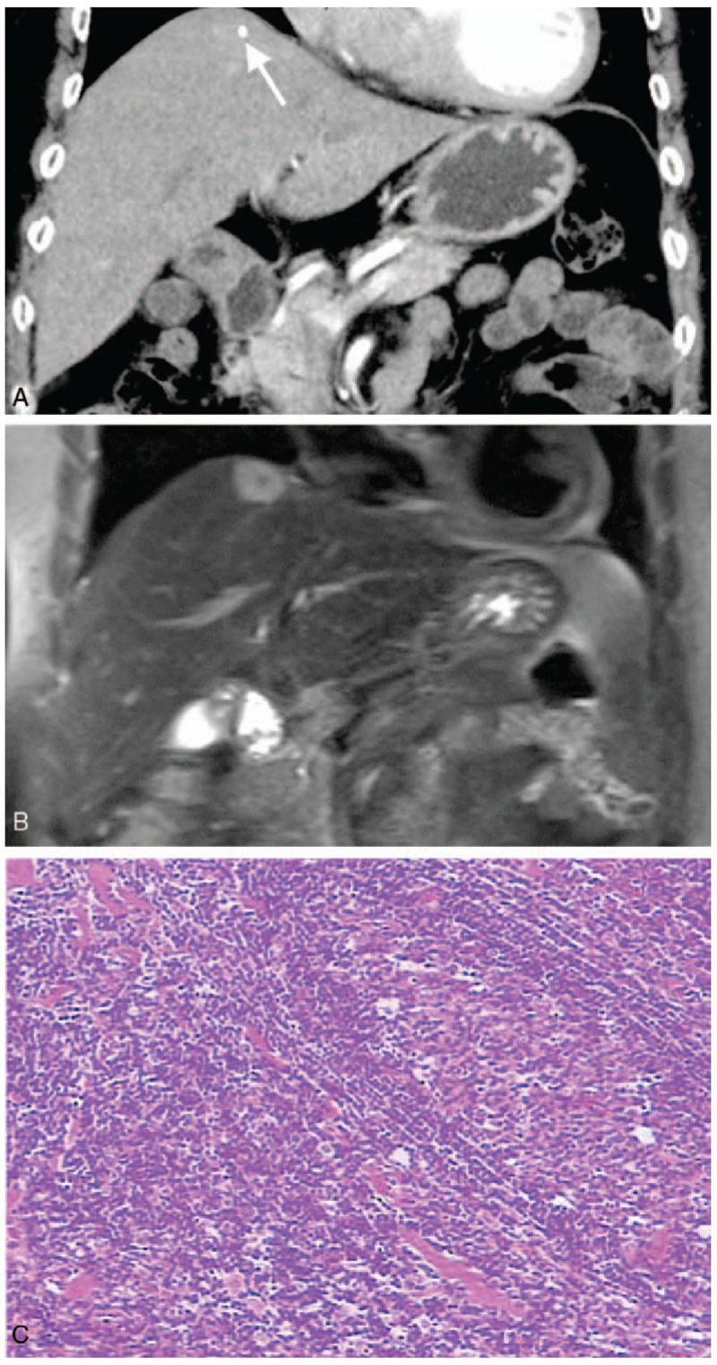
Figure 1.

Case 1: Hepatic CD in an asymptomatic 68-year-old woman. Coronal CT showed continuous enhancement of hepatic nodules at the parenchymal stage (A), with punctate calcification in the lesion (arrow); coronal T2WI showed hepatic nodules with hyper-signal and punctate hypo-signal (B); pathology showed a large number of lymphocytes arranged in concentric circles (C, HE × 200).
