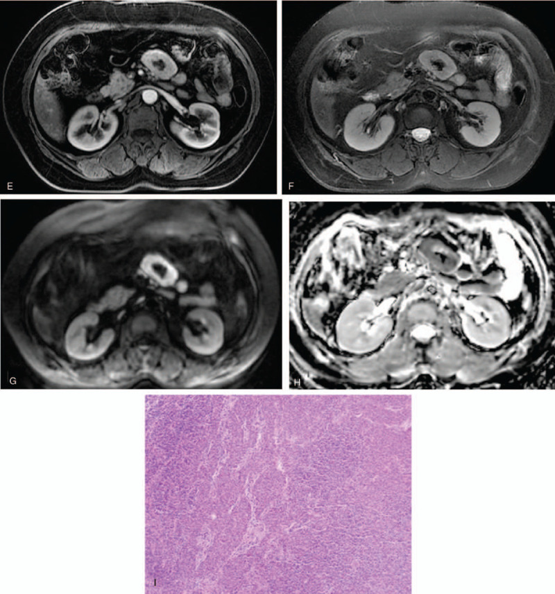
Figure 3 (Continued).

Case 2: Perimesenteric CD of CT and MRI findings in an asymptomatic 63-year-old woman. Unenhanced CT showed a perimesenteric mass with nodular calcification in the abdomen (A). Contrast-enhanced CT showed arterial enhancement of the mass, feeding vessel (B, arrow) could be seen in the lesion, and continuous enhancement at the delayed phase (C). T1WI showed iso/hypo-signal (D), T2WI showed mild hyper-signal, contrast-enhanced MRI showed the mass with an obvious enhancement at the arterial phase (E) and continuous enhancement at the delayed phase (F). DWI showed a hyper-signal region (G) and the ADC value was low (H). Pathology showed a large number of lymphocytes (I, HE × 200).
