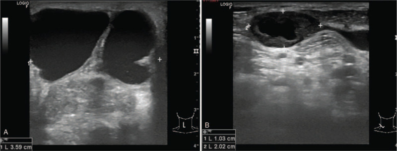
Figure 1.

(A) Neck ultrasonography at the last presentation showed heterogeneous echogenicity of solid lesion with multiple internal calcifications at right thyroid bed (size 2.1 × 1.6 × 2.8 cm with adjacent complex multilocular cystic lesion with septations and (B) enlarged right cervical lymph node with absent fat hilum.
