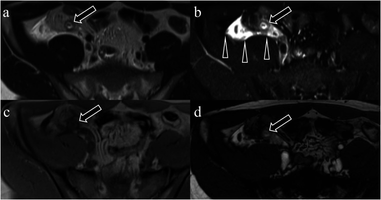
Fig. 3.
An 18-year-old man complaining of right lower abdominal pain diagnosed with acute appendicitis. (a) T1-weighted image. (b) T2W image. (c) FS-T2W image. (d) True-fast imaging of steady-state precession. Thickening of the appendiceal wall (intramural finding) is observed in all sequences (a–d, arrows). Fat stranding surrounding the appendix (extramural findings) is the most obvious on the FS-T2W image (b, arrowheads). FS, fat-suppressed; T2W, T2-weighted.

