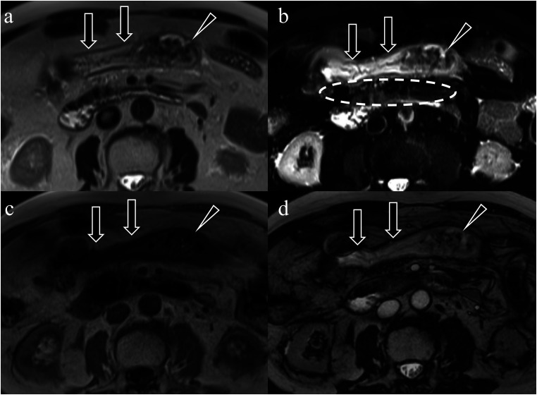
Fig. 4.
An 80-year-old man who presented with acute abdominal pain after defecation was diagnosed with necrotizing ischemic colitis. (a) T1-weighted image. (b) T2W image. (c) FS-T2W image. (d) True-fast imaging of steady-state precession. Intramural findings, including wall thickening (a–d, arrows) and the partial mucosal defect (intramural findings) (a–d, arrowheads) are observed in the transverse colon in all sequences. Mild fat stranding meaning extramural finding is observed in the transverse mesocolon in the FS-T2W image (extramural findings) (b, circle). FS, fat-suppressed; T2W, T2-weighted.

