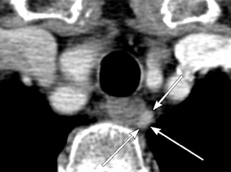
Figure 5d:

Images in a 69-year-old woman with primary hyperparathyroidism. Superior localization of four-dimensional (4D) CT compared with sestamibi SPECT/CT in a patient with multigland disease. (a) Axial delayed-phase sestamibi SPECT/CT shows focal sestamibi uptake localizing to a single abnormal left lower parathyroid gland (arrow). No additional abnormal focus is identified to suggest an additional abnormal parathyroid gland. (b) Axial contrast-enhanced arterial phase 4D CT shows enhanced left lower parathyroid gland (arrows) corresponding to same gland identified at sestamibi SPECT/CT. However, additional (c) enhanced right upper (arrow) and (d) left upper (arrows) parathyroid glands were identified at 4D CT, which were not observed at sestamibi SPECT/CT. The patient underwent bilateral four-gland exploration for multigland disease involving all three parathyroid glands.
