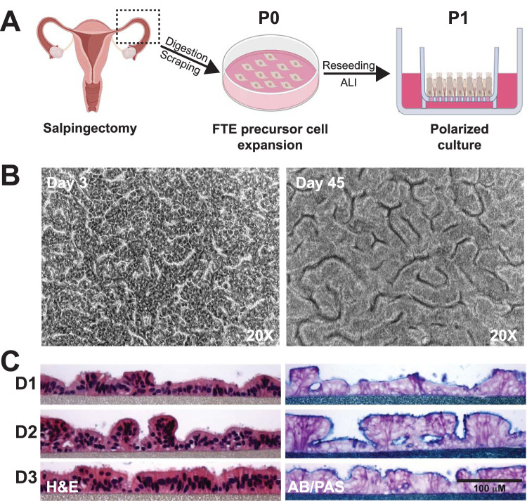
FIG 1.
Primary human fallopian tube epithelial (FTE) cells cultured in an air-liquid interface (ALI) polarize and secrete mucins. (A) Simplified model of the procedure to generate primary human FTE cell cultures. Fallopian tubes were obtained from women undergoing elective salpingectomies. Fallopian tubes were opened, diced into pieces, and enzymatically digested, and epithelial cells were obtained by gentle scraping of the lumen. Primary passage (P0) cells were then expanded on collagen-coated tissue culture dishes in a growth factor-rich medium, followed by dissociation and seeding of P1 cells on inserts or cryopreservation for later use. Creation of an ALI was induced on inserts by removing medium from the apical surface, which promoted epithelial polarization and cell differentiation. Images created with BioRender. (B) Cobblestone-like structures in FTE cell cultures were observed by phase-contrast light microscopy 3 days post-ALI, which was maintained through 45 days post-ALI. (C) Columnar polarized multiciliated and secretory cells were observed by hematoxylin and eosin (H&E) staining, and mucopolysaccharides were observed by alcian blue and periodic acid-Schiff (AB/PAS) staining of histological sections of FTE cell cultures from 3 donors 23 days post-ALI.

